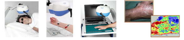
The PeriScan System is a Laser Doppler Perfusion Imaging system for non-invasive imaging of superficial tissue blood perfusion. It is operated from a standard PC and the patented scanning technique offers excellent image integrity, sensitivity and perfusion baseline stability. The technique can be used to monitor microcirculatory activity in healthy and diseased tissue, and show basal values and also responses resulting from an applied physiological stimulus, a "provocation".
Based on the Doppler principle, two-dimensional maps (imaging mode) or time traces (monitoring mode) of the tissue blood perfusion can easily be created. Easy-to-use image analysis software (LDPIwin) assists in the evaluation of the results and in report generation. The patented stepwise laser beam movement makes it possible to read low perfused areas.
Since no physical contact with the tissue is necessary, and no dyes or tracer elements are used, the influence on the perfusion can be kept to a minimum. These features also imply that repeated clinical investigations of e.g. healing wounds and leg ulcers can be performed without the additional risk of contamination, infection or discomfort to the patient.
The PeriScan PIM 3 System is flexible, small and of low weight which makes it easy to handle. The scanner head is directly connected to two USB2.0 ports in a stationary or laptop computer. A low power laser(Class 2) means that no additional safety precautons are required. The PIM 3 System is equipped with a digital camera for easier orientation in the images.
Applications
The PeriScan PIM 3 System can be used for numerous different types of applications, both in clinical and in research environment. See the PeriScan Literature Reference list at www.perimed.se or search the PubMed database www.pubmed.com.
- Angiogenesis and Growth Factor
- Darmatology
- Diabetes
- Raynauds Syndrome
- Wound Healing
- Visceral Surgery
- Brain
- Nerves
- Burns, Plastic Surgery and Transplantations
- Iontophoresis
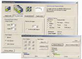
LDPIwin software
The LDPIwin software runs on a PC and is used to control the PeriScan PIM 3 System and assists in evaluation. With the software it is easy to set up shape, resolution ans size of the preferred measuring area.
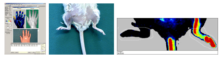
Imaging mode The perfusion over the area can be presented with its numerical values as well as a color coded image. In the user defined regions of interest(ROI) the software calculates parameters such as mean, maximun, minimum, % change, number of measurement sites and other values. If desired, the calculations and raw data can be exported to other programs(e.g. Excel, Word) for further evaluation.
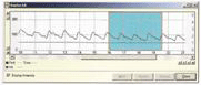
Monitoring mode In monitoring mode(also called duplex mode). Temporal variatons of the tissue blood perfusion in a single site can be recorded. The LDPIwin software assists in evaluation and calculations.